With the combination of natural inorganic elements, Osteobone can promote the proliferation of human bone cells, the function of bone morphogenetic protein, and the growth of new bone.
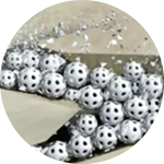
It adopts leading 3D printing technology to build biological micro-structure, which guarantees the access, proliferation and differentiation of bone cells, growth of new vessels, and exchange of metabolite. Furthermore, Osteobone has best matching rate between material degradation and new bone growth.
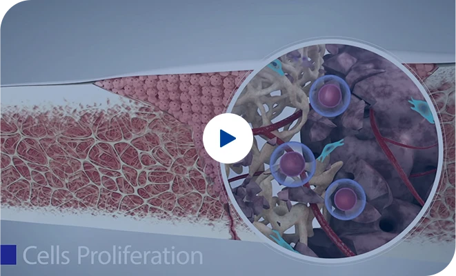
Osteobone is a safe and effective artificial bone product with three key advantages: osteoinduction, bionic 3D structure, and optimal material degradation and new bone growth balance. Suitable for non-weight bearing areas, it is used in orthopedic trauma, spinal fusion, and maxillofacial surgery. When applied, Osteobone granules should be mixed with blood and covered by periosteum or an artificial collagen membrane. The scaffold, comprised of silicon, calcium, phosphorus, oxygen, magnesium, and sodium, supports bone stem cells and undifferentiated mesenchymal cells to grow, eventually integrating with the original bone. Osteobone's success is due to ideal bone cell growth space, necessary blood vessels, interconnected scaffolds, and metabolite excretion channels.
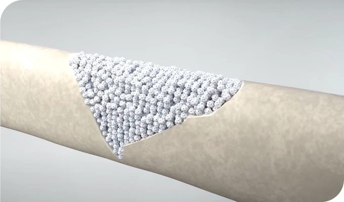
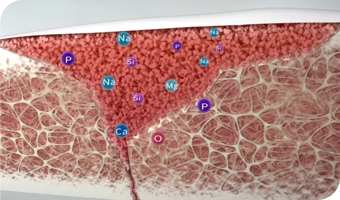
It is indicated for the repair treatment of defective bone tissue in non-weight-bearing parts of the human body such as orthopedic trauma, spinal fusion, maxillofacial surgery.
 Orthopedic Trauma
 Spinal Fusion
 Maxillofacial Surgery
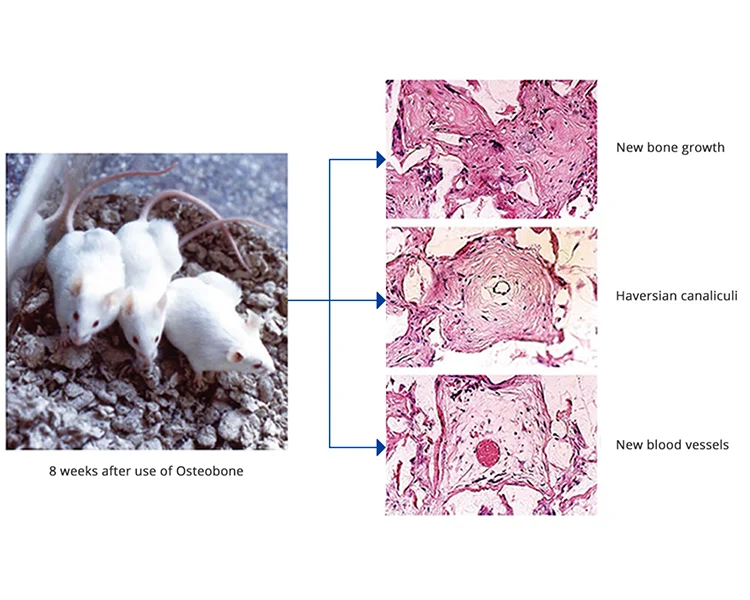
Following the use of Osteobone in laboratory animals, significant progress was observed after an eight-week period. The treatment led to the emergence of new bone growth, indicating successful bone regeneration and repair. Additionally, the formation of Haversian canaliculi, which are microscopic channels within the bone matrix, suggests improved bone structure and organization. Furthermore, the development of new blood vessels in the treated area points to enhanced vascularization, a critical factor in the bone healing process. Overall, these outcomes demonstrate the effectiveness of Osteobone in promoting bone recovery and restoration in experimental animals.
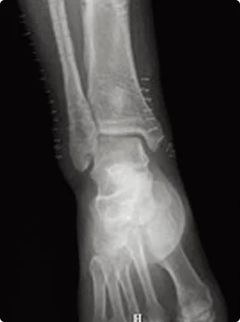
Two cases of bone defect after the excision of femur tumor were treated by Osteobone material and control group material respectively. 24 weeks after the surgery, it showed that Osteobone material had completely degraded and the new bone had come through. While the implant material of control group had not degraded, which blocked the repair of new bone.
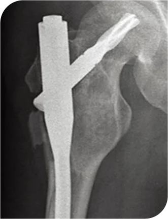
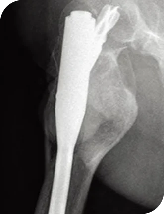
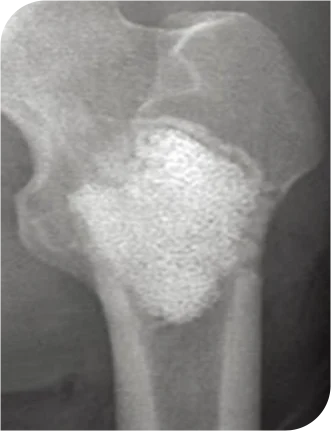
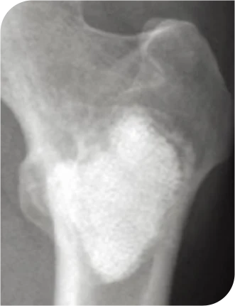
Two cases of bone defect in posterior superior iliac spine for bone transplatation were treated by Osteobone and control group material respectively. 24 weeks after the surgery, it showed that Osteobone material had completely degraded and the new bone had come through. While the implant material of control group had not degraded, which blocked the repair of new bone.
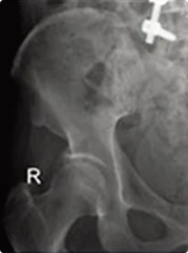
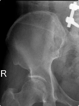
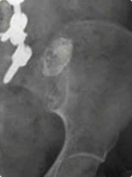
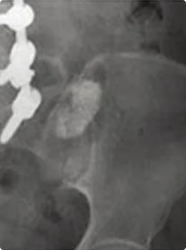
Two cases of bone defect after taking out inner bone nail were treated by Osteobone and control group material respectively. 24 weeks after the surgery, it showed that Osteobone material had completely degraded and the new bone had come through. While the implant material of control group had not degraded, which blocked the repair of new bone.
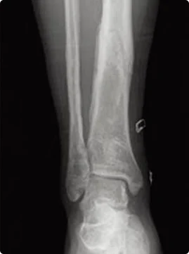
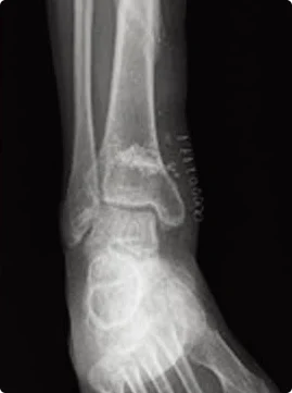
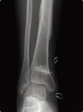
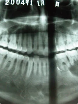
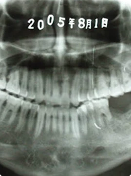
Bone defect after the excision of left side of mandibular cyst was treated by Osteobone. It showed that the new bone growed better 24 weeks after the surgery.
If you have any questions Please Email Us