is to provide excellent products to human being with
guaranteed quality and make the life be healthy and joyful.

2025-07-21


2025-07-05
Are you looking for the most effective wound care products? Do you know the advantages of Dermlin products? I will recommend Dermlin as the best wound care supplies and I will introduce the types, features, advantages, benefits and usage of Dermlin.
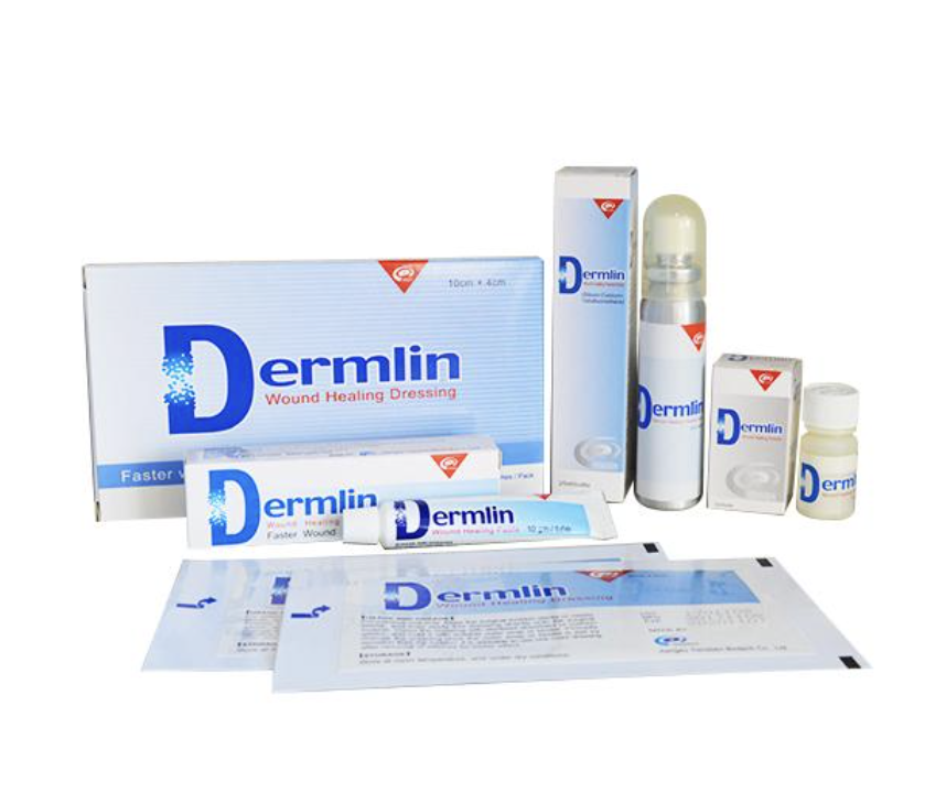
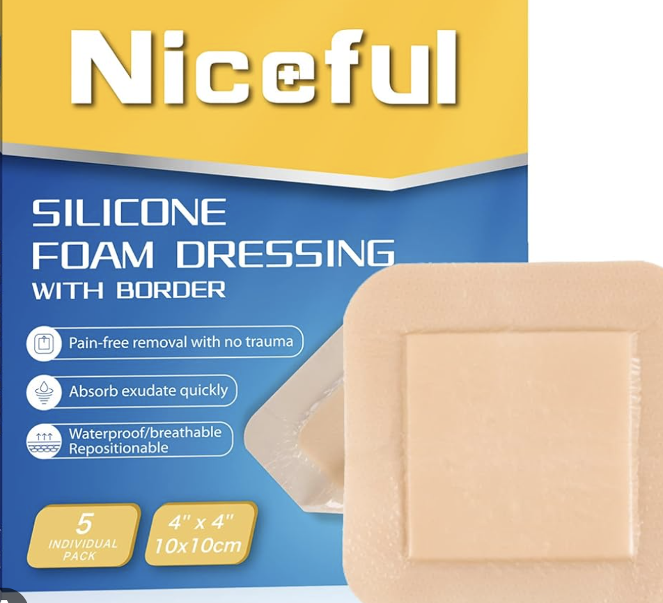
Dermlin is a wound care product specially in helping treat chronic and acute wounds.Dermlin Wound healing series contain four forms, powder, paste, powder spray and dressing. The main ingredients of these four forms are the same, consisting of inorganic elements, like silicon, calcium. For technical and clinical sides, the four forms have no differences. The ingredients of products have been certification by ISO and tested by professionally medical institutions, help in wound care healing and effective wound care.
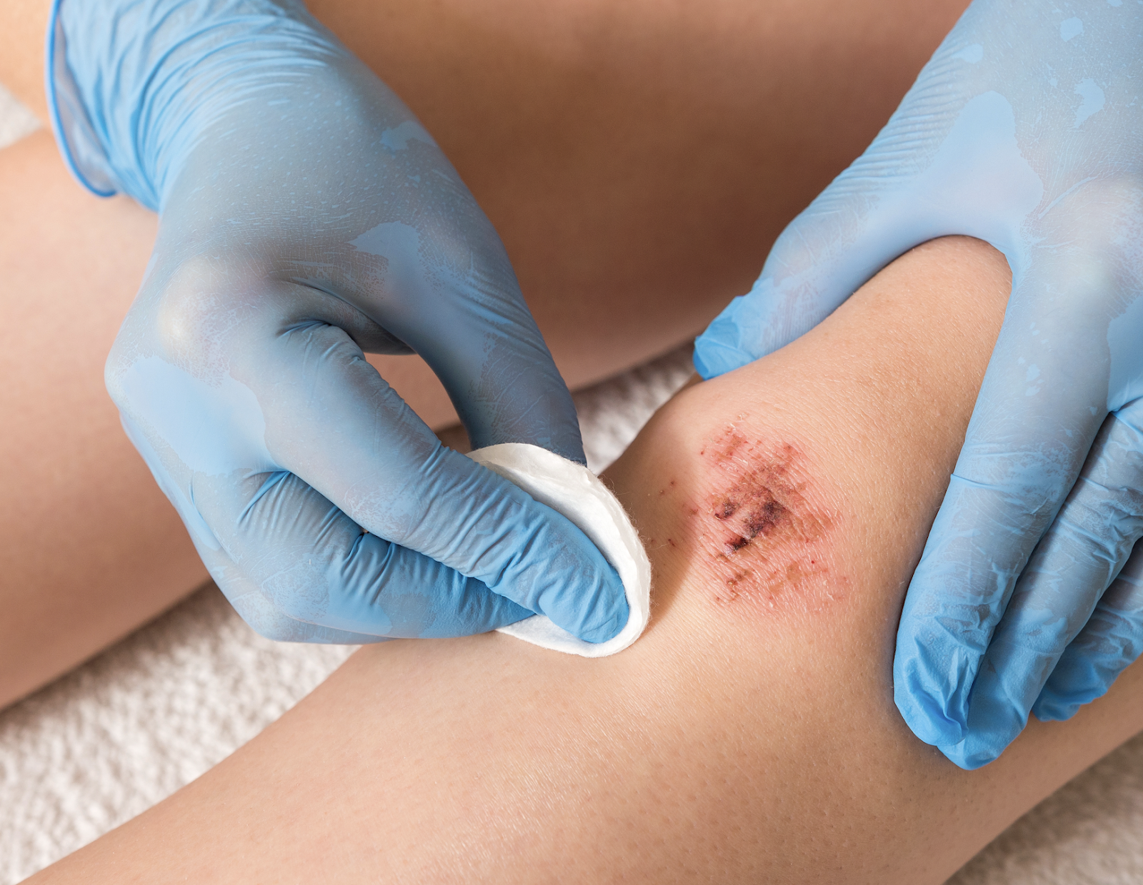
Dermlin can deal with different types of wound, whether it is a minor injury or a chronic wound and acute wound, Dermlin can quickly stop bleeding, promote wound healing, prevent infection, and manage scars.
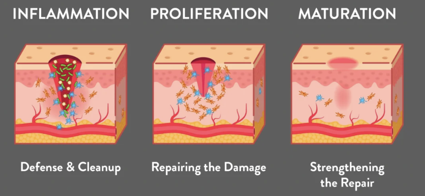
A chronic wound is a wound that does not progress through the normal stages of wound healing—haemostasis, inflammation, proliferation, and remodeling—in a predictable and timely manner. Typically, wounds that do not heal within three months are classified as chronic.Common chronic wounds include pressure ulcers, diabetic foot ulcers, venous ulcers, etc. Dermlin can manage different chronic wounds and patients’ health effectively.
Dermlin dressings absorb exudate and promote rapid healing so that reduce the risk of of infection.
Diabetes lead to ulcers, and if these ulcers are not well cared for and treated, there is a risk of amputation. In order to lower the risk of amputation, Dermlin dressings can effectively treat ulcer wounds, thereby promoting wound healing.
Dermlin absorb exudate,promote rapid healing and reducing the risk of recurrence.
An acute wound is an injury to the skin that occurs suddenly, rather than over time. It heals at an expected rate that is predictable by the normal wound healing process. Common acute wounds include burn wounds, traumatic wounds and surgical wounds, etc. Dermlin can manage different acute wounds and patients’ health effectively.
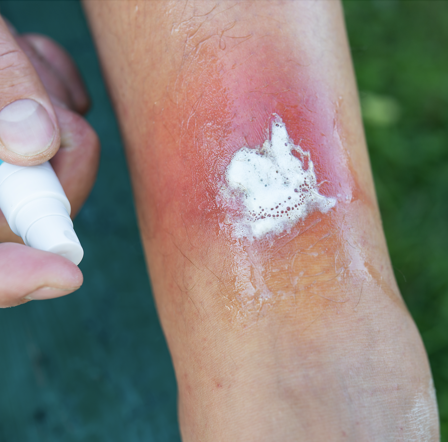
Dermlin can treat large burn wounds, promote epithelial tissue regeneration and healing, while reducing scars and achieving better cosmetic results.
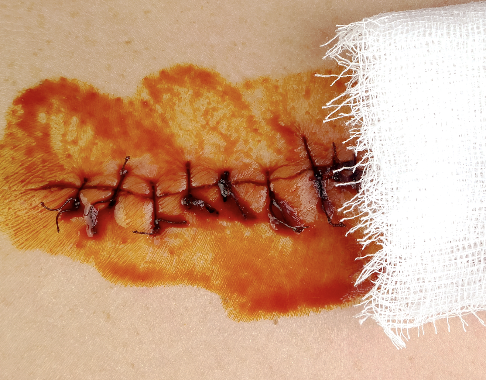
For various traumatic wounds, Dermlin dressings can quickly stop bleeding, resist infection and promote healing.
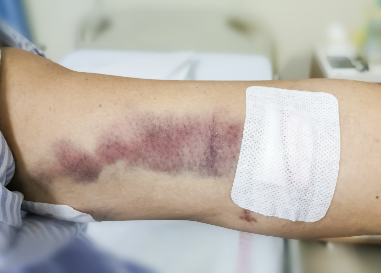
Dermlin dressings absorb exudate, resist infection, and promote rapid healing, reducing the risk of complications and improving postoperative outcomes.
Miner Injuries include cuts, scratches and scrapes, Dermlin dressings promote hemostasis, absorb exudate and promote rapid healing, meanwhile, it also has significant anti-inflammatory effect, so that preventing wound infection.
There are four types of Dermlin series products,every types of products have advantages and disadvantages and fits different type of wound. All Dermlin types products all satisfy you different need of wound management.
The main ingredient is calcium, which can quickly stop bleeding and reduce scar formation. It is suitable for minor wounds and diabetic ulcers, but the powder needs to be fixed with gauze. Dressings need to be changed regularly to prevent wound infection.
The core ingredient is calcium and silicon. It can provide long-term moisturizing for wound, so that promoting wound healing, regulating the synthesis of collagen and reducing the the formation of scars. It has the best anti-infection effect, which help in the recovery of chronic wounds and postoperative wounds better. In order to avoid the risk of allergy, conducting an allergy test before use to check whether you are allergic to the material of Dermlin dressings.
The core ingredient is a suspension powder, which can not only promote wound healing, but also effectively fight inflammation and inhibit bacteria. It is easy to carry and can be used in any occasion.
Ointment has a cream texture and contains inorganic elements such as calcium and silicon. It can reduce wound healing time and has the best effect on the care of chronic wounds, but its anti-infection effect is limited.
Dermlin become the most popular wound care product with an outstanding reputation base on the uniqueness and benefits that include innovative technology, high security, multi-function, wide application range, economy and convenience of Dermlin.
Dermlin series products use a combination of inorganic elements, with the core ingredients including calcium and silicon. In addition, its ingredients have been strictly verified for safety and have passed ISO verification, with no adverse reactions found, it suitable for long-term use.
Many research indicate that Dermlin series products have significant role in reducing the time for wound healing. For example, ithe healing time of the Dermlin group was shortened by about 10 days compared with the control group during the treatment of diabetic ulcers.
Dermlin products have the function of rapid hemostasis and anti-inflammatory properties, which can reduce inflammatory responses, promote tissue repair and effectively prevent the occurrence of secondary infections.
The Dermlin series of products regulates the synthesis and secretion of collagen, promoting wound healing while reducing scar formation, thereby improving wound appearance and patients’ satisfaction.
Dermlin series products are suitable for various types of skin wounds, including burns, abrasions, diabetic ulcers, bedsores, etc. Its versatility and wide applicability make it an important choice in clinical treatment.
Dermlin series products are cost-effective and effectively reduce medical costs. They are easy to store and use, and do not require refrigeration, making it convenient for medical staff to perform treatment in various environments.
Correct and safe use of Dermlin products can help Dermlin's healing effects on wounds.
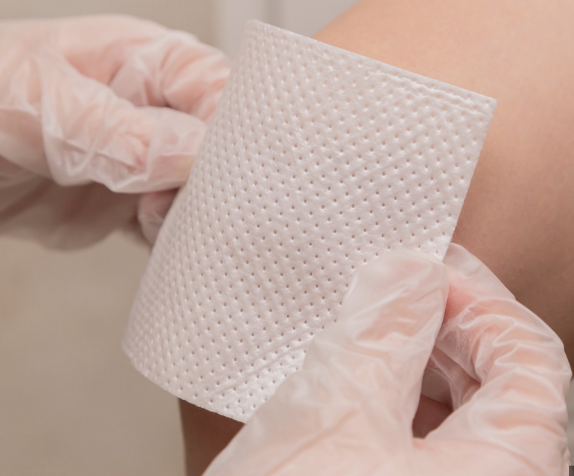
1. Completely clean and disinfect the wound, including removing the necrotic tissue.
2. Check whether patients are allergic to Dermlin products.
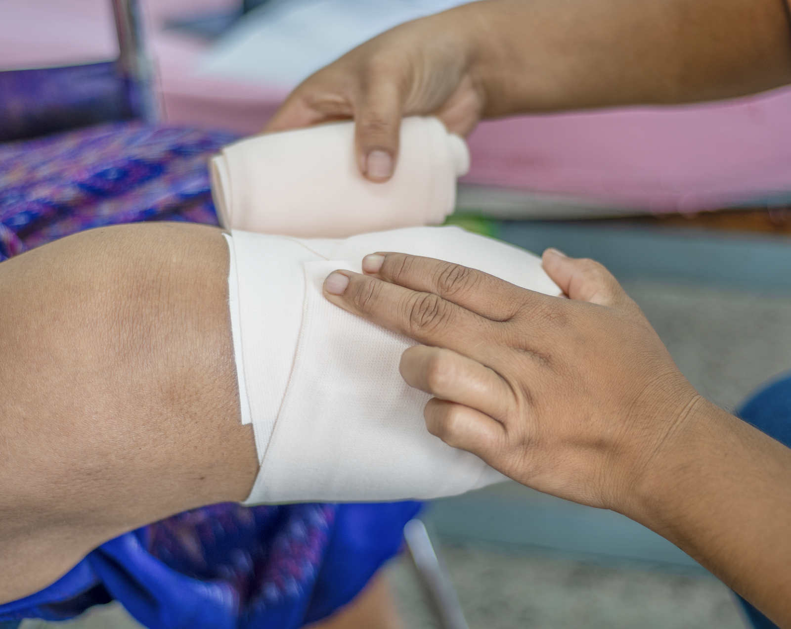
3. Apply a thin and even layer of Dermlin on the wound. And using gauze or other tools to fix the wound in according with the needs of wound care.
4. Change Dermlin once a day or two days according to wound condition.
Dermlin is a safe, efficient and widely applicable wound care solution. Through its unique technology, Dermlin wound care products can not only effectively promote wound healing and reduce scar formation, but also significantly shorten healing time and reduce the risk of infection.

2025-05-28
Wounds and blisters that appear inside the mouth are called mouth ulcers. They are painful and cause discomfort in a person's normal living. Eating and drinking become too difficult due to pain and irritation in the mouth. The pain sometimes becomes unbearable, and the person feels unable to speak properly and this requires the needs of the best mouth ulcer treatment.
Mouth ulcers are not rare cases; they are normal and can heal naturally within 1 or 2 weeks, depending on the condition. Despite its natural healing process, the things that make it difficult to bear are pain and irritation.
Two treatments for soothing the sores and speeding the healing process are oral ulcer patches and oral ulcer sprays. Both products offer several advantages, like reducing pain and discomfort by providing a soothing effect, but which one works most efficiently needs a comprehensive comparison. First, we'll discuss mouth ulcers briefly to understand which healing companion will win in the war: oral ulcer patch vs oral ulcer spray.

The mouth has a delicate tissue lining which is known as a mucous membrane. Any damage to this outer lining or erosion of a portion of it is a canker sore. Harm and injury to the mucous membrane mostly occurs accidentally. Ulcers need only 10 to 14 days for natural healing. They are painful and irritating but are not harmful like other diseases. If your ulcer doesn’t heal within 2 weeks or after medication, maybe it’s a symptom of mouth cancer.
There are various factors, from accidental biting to autoimmune condition, which causes mouth ulcers; a few are mentioned here.

It is a medicated, sterile adhesive film or gel patch used to apply on oral ulcers and treat them. These patches come with a properly packed style in aseptic packaging. They are also called Canker sores, and they appear on the gums of mouth, on and undersurface of the tongue, and cheeks inside. Oral Ulcer Patch works by following mechanisms:
It covers the oral ulcers and protects them from the material coming inside the mouth like food particles and drink. It works as a helpful barrier to mouth sores against outside material and saliva.
It relieves the pain of an ulcer because most of the patches contain ingredients for numbness and soothing.
These patches promote the healing process because they contain hyaluronic acid, aloe vera, and antiseptic material.
It prevents the spreading of ulcers in the mouth by covering them and keeping them stuck in the initial place. It helps prevent further irritation and potentially reduce the risk of spreading if the cause is contagious (though canker sores themselves are not typically considered contagious)
Note: The ingredients for numbness and healing process vary from brand to brand; the mentioned ones are common but not available in all products.
Improper placement may cause patches to fall off easily
Some sores appear in difficult-to-reach areas in the mouth where applying an oral ulcer patch is hard.
Some people may feel foreign body sensations and uncomfortable feelings inside their mouths.
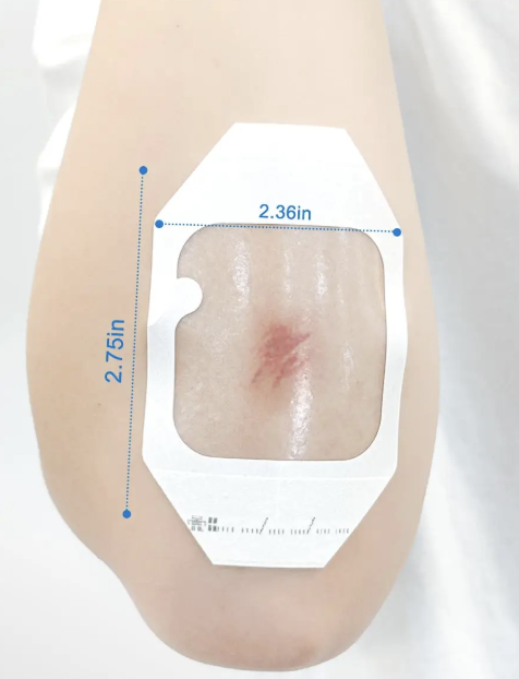
First, clean and dry the area around the ulcers with an aseptic cloth or gauze piece. Then, apply the patch on the ulcer specified gently and keep pressing for a while so that it is fixed properly. Most likely, it's an easier way to use it, but sometimes it causes foreign body sensations in the mouth.

Mouth ulcer spray is a medicated solution in liquid form made for oral use to treat oral ulcers or canker sores. It usually comes in a spray bottle and is very easy to use in out-of-reach areas in the mouth. It contains the following ingredients:
Note: not all oral ulcer sprays will contain all of these ingredients. Different formulations may prioritize different mechanisms of action. For example, some might focus more on pain relief, while others emphasize antiseptic properties or healing.
The process of using mouth ulcer spray to speed up the canker sores healing process is easy and quick. Clean the area of the ulcer with clean gauze or tissue paper if possible. Take a spray bottle in one hand, open your mouth in front of the mirror, and spray directly on the ulcer area. This spray can be used two to three times a day or as directed by the physician. It’s better to not eat or drink anything within 30 minutes after spraying.
Both oral ulcer patches and oral ulcer sprays have some pros and cons, but the best option lies in their need and the location of the ulcer.
Oral ulcer spray comes with easy applications in which a person needs to spray the liquid on the sore directly. An oral ulcer patch is a solid product that users can apply on the mouth ulcer with careful handling. It may cause pain due to pressing the patch over sores. Wrong placement may cause an oral ulcer sheet to fall off while eating or talking. From an ease in applying point of view, spray is better for one or more sores than patches.
Both patches and spray can only speed up the natural healing process by reducing pain and discomfort. Patch offers relief slowly, which benefits in the long term, while spray offers quick results, but it fades after some time due to saliva and other pollutants. The long-term benefits make oral ulcer patches a better option than spray.
An oral ulcer patch becomes a soft gel after reacting with saliva and keeps its place for long hours. It has no bad taste and doesn't cause a burning sensation inside the mouth. Oral ulcer spray can easily wash away due to its liquid nature. They have a bad taste which becomes difficult for a person to bear. So, here patches work better than spray. .
An oral ulcer patch is a small sheet that doesn't allow anything to come into contact with the mouth sore. The gel it forms after application works as a physical barrier against outside material, like food, drinks, and saliva. Oral ulcer spray is liquid that doesn't remain at the target area for a longer period and never works as a physical barrier for sores. The oral ulcer patch is better to choose here as a winner.
For a long-term effect, the choice will be an oral Ulcer Patch. Most of the time, these oral ulcers are so resistant that they are hard to treat. They persist for a long time, so for them oral ulcer patch works best. They adhere to the ulcer area and stay for hours at the target area.
Oral ulcer spray is short-lived and also easily washed away, so its effect on ulcers is not long-lasting, and most of the time, it remains ineffective, and ulcers stay as they are.
Oral ulcer spray can easily cover the mouth sore of whatever size. Each sore needs a separate patch, which is okay. Spray can cover the sore but for a short time. It also covers many wounds at a time, even those that appear in hard-to-reach areas.
Patches stick to the mouth ulcer and remain at their place for hours due to their sticking ability. They can cope with normal drinking and eating and don't affect the normal routine of a person. Liquid sprays just retain their position for a few minutes and then wash away, even because of saliva. Patches work for longer due to their sticking quality.
Oral ulcer spray doesn't require reapplication several times as it sticks to the targeted area for a longer period than sprays. You need to reapply spray 2 to 3 times a day because of its quickly dissolving nature.
Sores demand a gentle healing process because of associated pain and discomfort. An oral ulcer patch doesn't only accelerate natural healing but also keeps the targeted area clean. It doesn't let the irritants and bacteria come in contact with the sores.
Spraying also kills germs and bacteria but doesn't provide a shielding effect. Too much respraying also causes side effects because of drying targeted areas. So, an oral ulcer patch helps in faster but long-lasting healing instead of giving quick but short-lived effects.
For better understanding of users and wise decisions Here is the quick comparison table of oral ulcer patch vs oral ulcer spray.
Oral ulcer patch wins in our comprehensive comparison, so they are the best mouth ulcer treatment for all who want an effective remedy. It offers a gentle treatment that reduces ulcer pain and irritation, which are the most trouble-making for a patient. Its easy application and pain-relieving effect make it the best mouth ulcer treatment than oral ulcer spray.
An oral ulcer patch is the best option for speeding the mouth ulcer healing process. Among so many ulcer patch options available in the market, Ulceloocin Oral Ulcer Patch is considered the best mouth ulcer treatment.
It has calcium, silicon, and phosphorus in its composition, which are natural minerals and help in the natural healing of the mouth sores. Its drug free nature helps in making them a better solution for all ages, even for children. It contains no hormones, which is why it's ideal to use for pregnant womens.
After applying to the mouth sore, the Ulceloocin turns into a soft gel after coming in contact with saliva. It covers the mouth ulcer and works as a physical barrier against everything from outside material to saliva. It offers a soothing effect and reduces pain and irritation. Then, it accelerates the healing process and cures your sore by helping mucous tissue grow back with less pain and no burning.
Among oral ulcer patches and sprays, patches are a better option because of their easy application and long-lasting pain-relieving effect. They make a protective physical barrier at the ulcer and save it from the irritants like food and drinks.
They accelerate the healing process and enhance comfort level until a complete cure. As they stay at the targeted area for hours, there is no need to reapply them again and again. Hence it reduces dosing frequency and becomes cost effective.
What works best really depends on your lifestyle and the kind of mouth sore you’re dealing with. If you’re someone with a busy routine and prefer something easy to use, the patch could be a great fit. For many, it's the most convenient way to handle a mouth ulcer without adding stress.

 EN
EN  ES
ES PT
PT SV
SV DE
DE TR
TR FR
FR JA
JA